Find your ideal sample storage tube: Tube Wizard
Blog posts
How just labeling tissue sample tubes is not a good enough approach
The identification methods of tissue samples vary among research facilities and clinical institutions. Some scientists and clinicians are labeling sample tubes with handwritten codes, and are identifying and tracking tissue samples on the basis of Excel spreadsheets. Other researchers combine printed labels with barcoded tubes to identify samples. Researchers worldwide have not reached a consensus about the best method of identifying tissue samples. Furthermore, researchers often have to consider resource constraints when choosing between different identification methods. It’s however safe to assume that no researcher intents to loose valuable tissue samples. Losing or mixing up tissue samples could lead to poor diagnostics, delaying drug discovery, postponing research or losing irreplaceable information.
Labeling tissue samples manually is a common practice. Handwritten codes are for example used as a low-cost and easy solution to identify samples. Nevertheless, there are risks with identifying and tracking tissue samples handwritten codes. Even the best staff member can mislabel a tube, leading to a lost sample. If the staff members write all code labels perfectly, there could be a problem with the identification of the code. The number ‘6’ could be mistaken for the number ‘8’, the number ‘7’ might be mistaken for the number ‘1’ etc. Furthermore, a research facility could switch between identification systems throughout the years (this typically happens when biobanks keep growing). After systems and staff members change, deviations from identification protocols might occur.
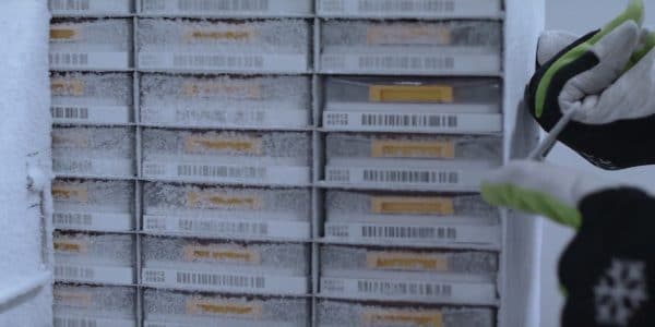
A printed label could also lead to sample loss. Many tissue storage tubes are used to store samples in extreme conditions. Consequently, it’s important that the label on a tube can withstand ultra-low temperatures. To be sure of this, labels should have been tested in the appropriate conditions. The challenging issue with applied labels is that the label can separate from the tissue tube inside the freezer, leading to unidentified specimens. Losing tissue samples is no small matter. Tissue samples are irreplaceable and thus invaluable. Moreover, there are safety concerns about having unidentified samples in a laboratory. After all, a sample might unknowingly contain infected tissue.
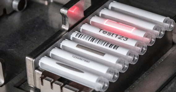
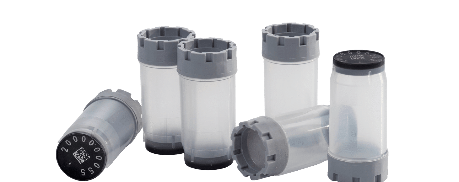
An alternative to handwritten or printed labels are laser-etched 2D codes. These codes are laser-etched into the black code surface on the bottom of the Micronic tissue tubes. The black code surface cannot be separated from the transparent tube due to a 2-component injection molding process that ensures that the code surface is fused as one to the tube. This makes it impossible to lose a sample due to a lost or mixed up label. Laser-etching a human-readable code on the tube allows you to visually identify a specimen without the possibility of illegible handwriting. 2D Data-Matrix coded tubes are used by many research laboratories worldwide in order to fully track and trace valuable samples during the entire sample storage process. The 2D code can be used to identify the sample, its coordinates within a storage rack, the particular rack and the location of the rack in the freezer. Information about the location and identity of each sample can be gathered by scanning the 2D Data-Matrix code of a single tube or a whole rack. A 2D Data-Matrix code scanner can be integrated with a Laboratory Sample Management System. This procedure of digitally gathering information ensures the complete traceability of tissue samples. Having a workflow with 2D coded tubes, code readers and the right software reduces the change that critical research has to be delayed or discontinued.
Newsletter
Subscribe to our newsletter
Be the first to be informed about our exclusive offers, the latest innovations, upcoming events and more!
Micronic Newsletter
The following articles might be of interest to you as well